


Cell Imaging
The Cell Imaging platform provides research teams with its expertise and a wide range of high-performance equipment for photonic and quantitative imaging, at low and high throughput.
Contact : jpolentes[AT]istem.fr
The Cell Imaging platform, whose members have worked for several years in research teams at I-Stem, puts its experience in particular in cell biology at the service of research teams inside or outside I-Stem. It can thus easily understand and respond to the researchers’ problems, either thanks to the equipment available on site, or by integrating or developing new tools on demand.
The platform, created in 2016, offers imaging techniques from the subcellular level to the whole organoid. It brings together a complete set of equipment grouped around several complementary areas of activity:
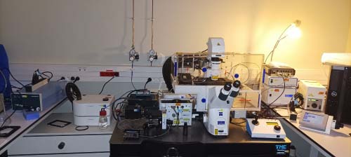
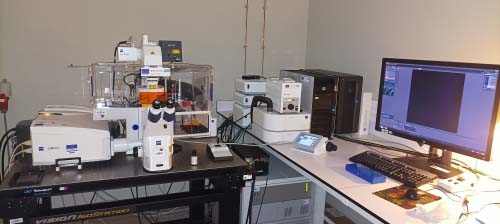
1- Photonic Imaging
The photonic imaging pole gathers equipments that allow the imaging of cells, tissues, organoids or embryos on different supports (slide, petri dish, multi-well culture plates, culture flasks). Different imaging modes are available, such as bright field imaging in color (Evox XL Core), epifluorescence (2 Zeiss Observer Z1), or high resolution confocal imaging (Zeiss spinning Disk microscope and Zeiss LSM-880 Airyscan). The equipment also allows to work on fixed or living samples, in standard 2D or multidimensional imaging (3D, live imaging, multiposition, in situ imaging).
Functional imaging studies (Calcium Flow Analysis, Optogenetics, FRAP) can be performed on high resolution microscopes.
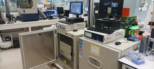
2- Quantitative automated imaging
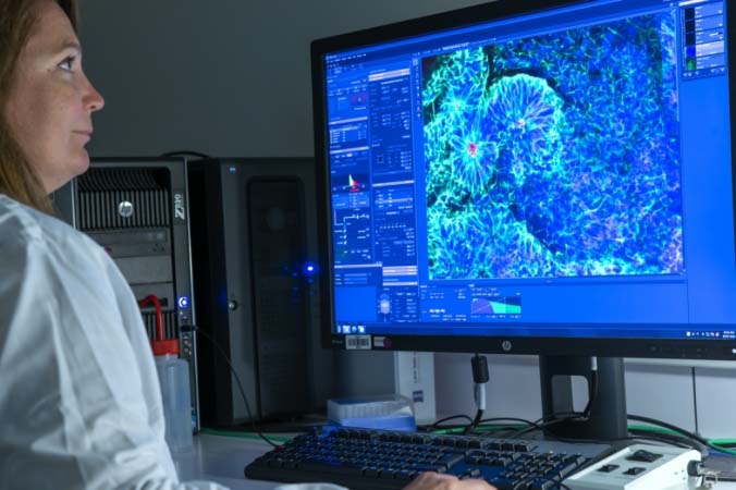
The Quantitative Imaging Center offers automated optical equipment that combines epifluorescence acquisition with high throughput and high content image analysis.
Three high-content imagers (Molecular Devices ImageXpress micro XL, Molecular Devices imageXpress HCS.ai, Cellinsight CX7) coupled to robotic arms allow to scan and analyze up to a dozen multi-well plates according to several possible analysis algorithms. These machines are compatible with pharmacological molecule screening studies.
One Incucytes imager (S3, Sartorius), installed in dedicated culture incubator, allow in situ monitoring of cell cultures by taking pictures at regular intervals over a period of up to several weeks. These pictures are then processed by quantitative analysis.
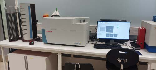
Finally, a plate reader FDSS µCell (Hamamatsu) allows a fast and dynamic acquisitionof calcium fluxes on one or more multi-well plates, coupled with an analysis of the intensity of the observed markings. This equipment is compatible with 96-well and 384-well culture formats, with the option to inject chemical inducers or inhibitors (injection heads) or to use electrical stimulation (96-well format only).
3- Analysis workstations
The platform is equipped with several computer analysis stations that complement the machines. In particular, it has a station equipped with IMARIS software for the optimal exploitation of images acquired in photonic imaging.
4- Technology watch, Research & Development
The members of the platform are always on the lookout for new technologies and analysis tools, in order to offer the best practical options to research teams. Our activity therefore includes a significant amount of research and development time in order to continuously improve the tools and services available.
In this approach, the Spinning Disk microscope has recently been updated to be compatible with 3D imaging of large samples (up to 1mm thick). The team’s skills now include these imaging techniques (cell culture of organoids, transparency treatments, 3D rendering and analysis of the images obtained).
Complementary image analyses are developed through software such as ImageJ, Metamorph, Imaris, Qupath, Cellpose, Stardist, or scripts developed in Python. The team is currently working on the integration of artificial intelligence in some complex analysis tools.
5- User experience on the Cell Imaging platform
The platform offers different ways of using the equipment depending on the volume of use and the comfort of the users: training users to use the equipment independently, or assisting research teams with complex image acquisition or analysis protocols.
Team members
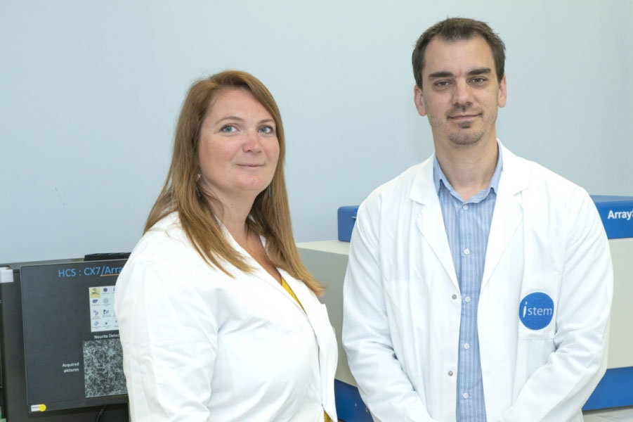
Jerome POLENTES
Platform Manager (CECS)
Jérôme joined I-Stem in 2008 as a research engineer and has been in charge of the platform since 2016. He is mainly in charge of photonic imaging and analysis tools development. Jérôme has a PhD in Neurophysiology & Neuroscience.
Céline LETEUR
Platform Engineer (CECS)
Céline has been part of the platform since 2016 after being part of the Neuroplasticity & Therapeutics team. Céline is mainly in charge of high content imaging and 3D microscopy development. Céline has a DEA in oncology.
Collaborations
Publications
High-throughput screening identifies bazedoxifene as a potential therapeutic for dysferlin-deficient limb girdle muscular dystrophy.
01 July 2025
British journal of pharmacology
MBNL-dependent impaired development within the neuromuscular system in myotonic dystrophy type 1.
01 February 2023
Neuropathology and applied neurobiology
CRISPR gene editing in pluripotent stem cells reveals the function of MBNL proteins during human in vitro myogenesis.
17 December 2021
Human molecular genetics
Evos XI CORE
Inverted bright field microscope equipped with a color camera for imaging stained histological sections (Funding: CECS)
Axio Observer Z1 (named respectively « Z2 » & « Z3 » )
2 inverted wide-field microscopes in epifluorescence or transmitted white light, equipped with various magnifications allowing them to work on different supports, with thin or thick-bottom support (Funding: Genopole)
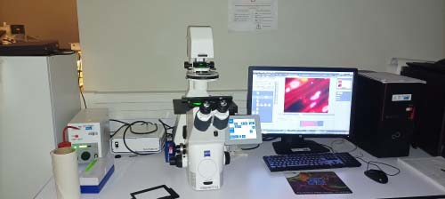
Spinning Disk
Versatile inverted microscope for fluorescence imaging in wide-field, or confocal in spinning disk mode. Compatible with imaging on living samples, and functional studies (Optogenetics, Calcium studies…). (Funding: Inserm; Genopole; CECS)
LMS880-Airyscan
Inverted confocal microscope for Fluorescent high resolution imaging of living or fixed biological samples fluorescence images. (Funding: CRCT)
Incucytes S3
Automated imagers installed in a dedicated culture incubator for monitoring cell cultures in situ. They can work on bright field or epifluorescence mode, and allow automated analysis of the images acquired. (Funding: Genopole, CECS; CRCT)
CX7
High content imager for multi-well cell culture plate screening, in epifluorescence or brightfield. The simple interface makes it possible to be quickly autonomous (Funding: INSERM)
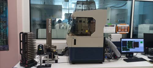
ImageXpress Micro XL
High content imager offering more possibilities than the CX7, including the possibility to work on live cells, to do volumetric analysis, and especially the possibility to create fully customized analysis algorithms. (Funding INSERM)
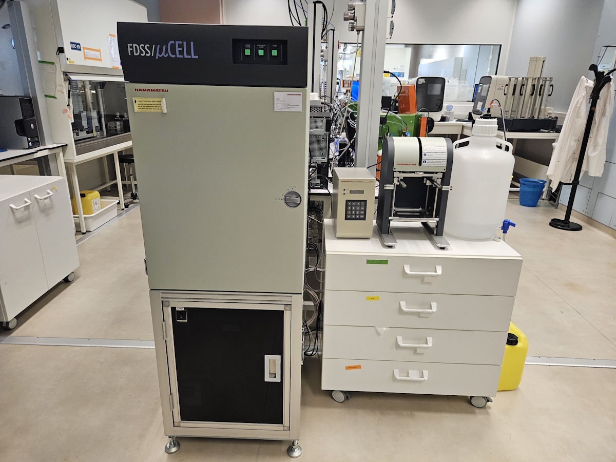
FDSS µCell
High throughput multi-well plate reader for fast, dynamic and low resolution analysis of intensity level variations of fluorescence or luminescence in biological labels. (Funding CECS)
Dedicated computer park
Several physical and virtual computer stations for image processing and analysis, with dedicated software or tools developed by the platform members.
– 1 computer equipped with IMARIS software (deconvolution, 3D rendering and image analysis)
– 2 computers dedicated to image analysis (ImageJ, Metamorph), to the consultation of Incucytes data
– 3 Computers+1 virtual machine (with MetaXpress software) for ImageXpress data analysis
– 2 virtual machines (with HCS Studio software suite) for the analysis of CX7 data
– 3 physical workstations equipped with In Carta software; for analyzing images from the HCS.ai imager by Molecular Devices
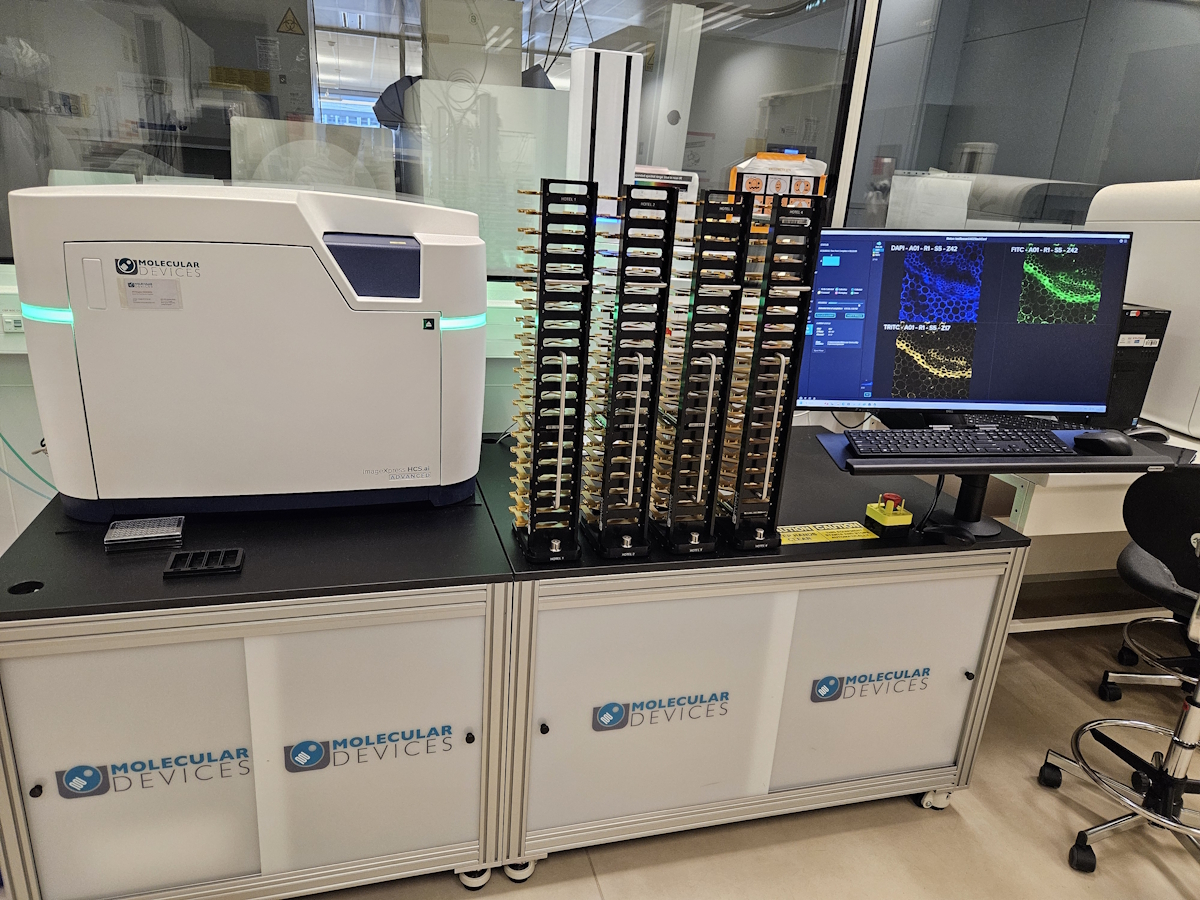
ImageXpress HCS.ai
State-of-the-art high-content imaging (HCS) system, enabling image acquisition and analysis on multi-well plates of all formats, compatible with brightfield, fluorescence, epifluorescence, or confocal imaging, on fixed or live samples, 2D or 3D, volumetric analysis, live monitoring, and tracking. Equipped with a robotic arm enabling batch analysis of multiple plates (compatible with high-throughput screening). (Funding Genother)